
Panoral Positioning Guide
Make sure your patients are correctly positioned
How you position your patient can dramatically affect the quality of your images. Compare your images to the examples below to help identify any common positioning mistakes to improve the quality of your images
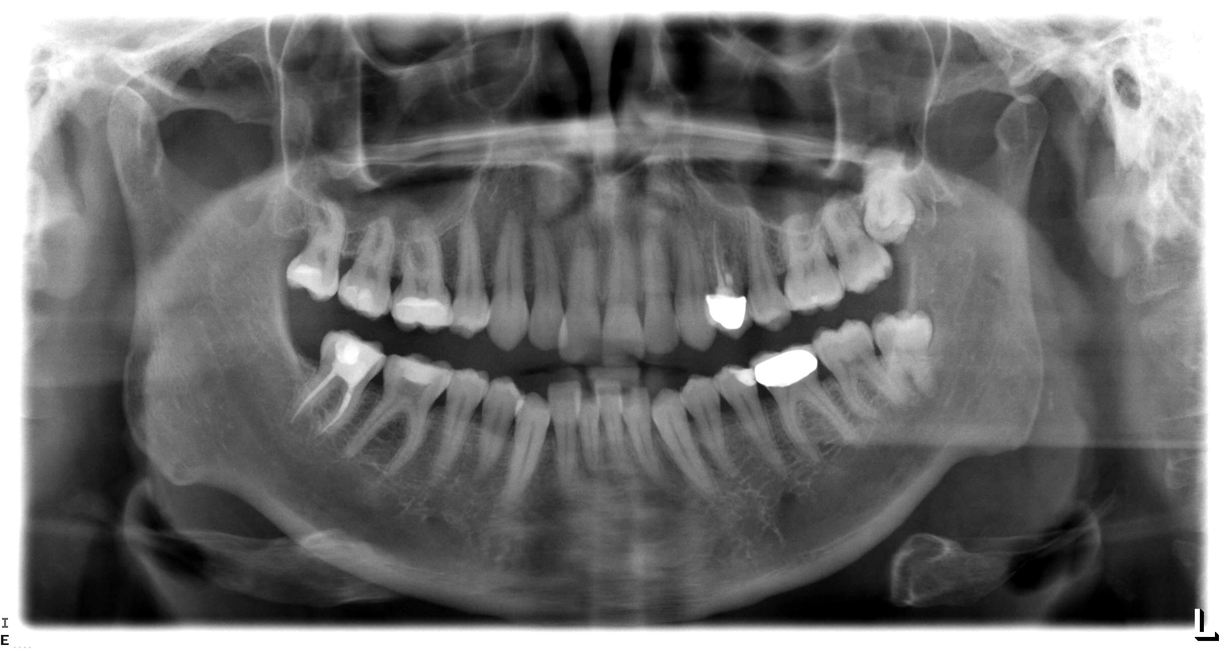
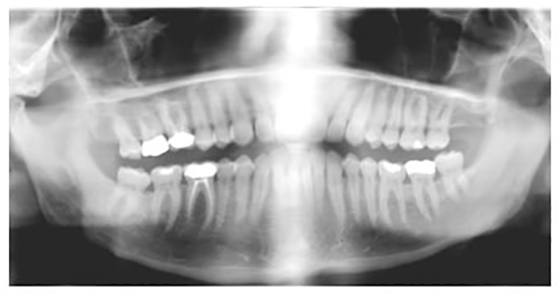
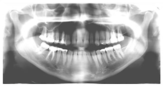
Good Example
This is an example of an image where the patient has been correctly positioned. You can see that in the image the teeth are aligned in a 'U' shape, almost as if they were smiling, and that the angles of the mandible are at the same height on both sides of the image as well as there being equal amounts of spine visible on both outer edges of the image.
The images below are all of the same subject, the only difference in each one being either the exposure setting or the position of the patient and as you can see they all have an effect on the image quality.
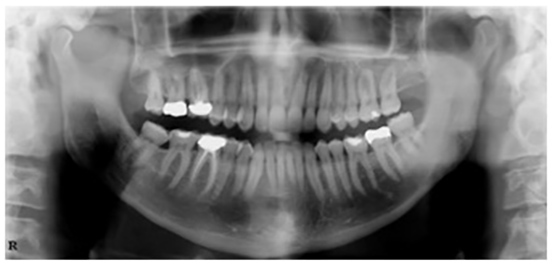
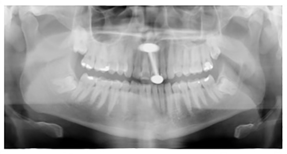
Patient too far Forward
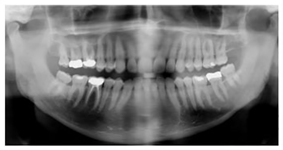
Patient too far Back
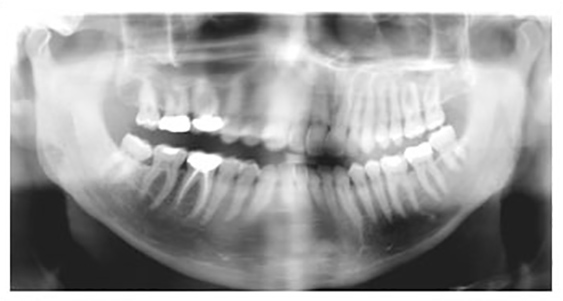
Patient not Centred
Ghost Artifact
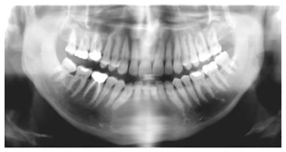
Chin too High
Chin too Low
Overexposed
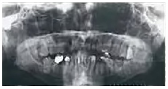
Underexposed
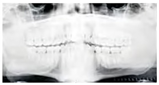
Tongue not in Roof of Mouth
 01283 246228